
Advancements in Automation and Technology in Healthcare: Revolutionizing Medical Labs and Phlebotomy
Summary
- Introduction of automation in medical labs
- Advancements in digital pathology
- Use of wearable technology in phlebotomy
Technology has revolutionized the healthcare industry, particularly in the field of medical labs and phlebotomy. Advancements in technology have significantly improved efficiency and accuracy in the analysis of tissue samples and blood draws in the United States. In this article, we will explore some of the key advancements that have transformed these areas of healthcare.
Automation in Medical Labs
One of the most significant advancements in medical labs is the introduction of automation. Automation has streamlined processes, reduced human error, and increased the speed at which tests can be conducted. In traditional medical labs, technicians would manually handle samples, perform tests, and record results. This manual process was not only time-consuming but also prone to errors.
With automation, robotics and advanced machinery can now handle a large portion of the testing process. This includes sample preparation, analysis, and result reporting. Automated systems can handle a high volume of samples simultaneously, allowing for quicker turnaround times and more efficient Workflow. In addition, automation reduces the risk of contamination and human error, leading to more accurate results.
- Automated sample processing: Automated systems can handle sample preparation, reducing the risk of contamination and ensuring consistency in Sample Handling.
- Robotic testing: Robots can perform a wide range of tests accurately and quickly, freeing up human technicians to focus on more complex tasks.
- Remote monitoring: Automation allows for remote monitoring of tests, enabling technicians to review results and make real-time decisions from anywhere.
Advancements in Digital Pathology
Digital pathology is another area that has seen significant advancements in recent years. Digital pathology involves the digitization of tissue samples for analysis, allowing for more efficient and accurate diagnosis of diseases. Traditional pathology involves the examination of tissue samples under a microscope, a process that can be time-consuming and prone to human error.
With digital pathology, tissue samples are scanned and converted into high-resolution digital images. These images can then be analyzed using computer algorithms, allowing for faster and more accurate diagnosis. Digital pathology also enables pathologists to collaborate remotely, share images for second opinions, and access a digital archive of past cases for reference.
- Whole slide imaging: Digital pathology systems can capture high-resolution images of entire tissue slides, allowing for detailed analysis without the need for a microscope.
- Image analysis algorithms: Computer algorithms can analyze digital images to identify patterns and abnormalities, assisting pathologists in making more accurate diagnoses.
- Remote collaboration: Digital pathology enables pathologists to collaborate remotely, share images, and consult with experts around the world in real-time.
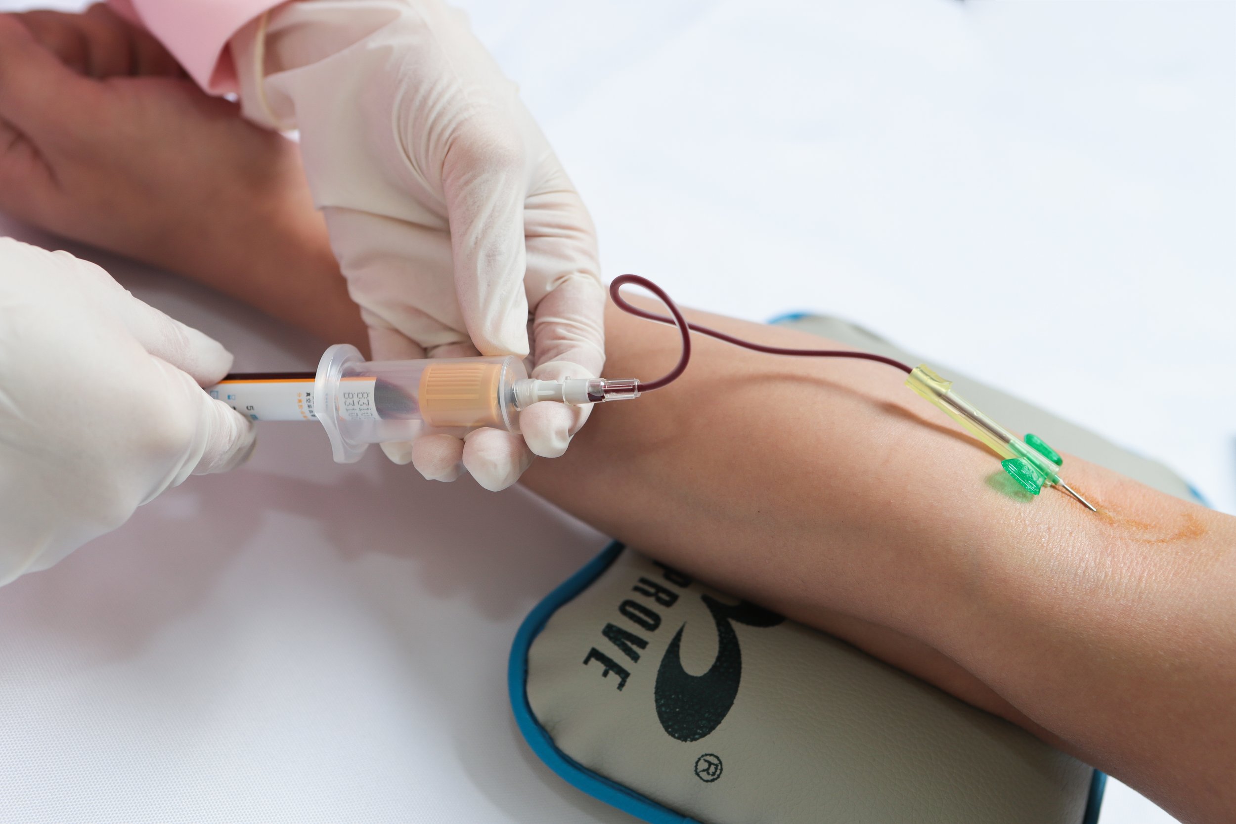
Use of Wearable Technology in Phlebotomy
Phlebotomy, the practice of drawing blood from patients for diagnostic testing, has also benefited from advancements in technology. One notable advancement is the use of wearable technology in phlebotomy procedures. Wearable devices such as smart glasses, smartwatches, and handheld devices have revolutionized the way blood draws are performed, improving efficiency and accuracy.
Wearable technology in phlebotomy can provide real-time guidance to phlebotomists, ensuring proper technique and reducing the risk of errors. These devices can also streamline documentation processes, allowing for quicker and more accurate recording of patient information. In addition, wearable technology can improve communication between phlebotomists and Healthcare Providers, leading to better coordination of care.
- Real-time guidance: Wearable devices can provide step-by-step instructions to phlebotomists, ensuring proper technique and reducing errors.
- Streamlined documentation: Wearable technology can streamline the documentation process, allowing for quicker and more accurate recording of patient information.
- Improved communication: Wearable devices facilitate communication between phlebotomists and Healthcare Providers, leading to better coordination of care and improved patient outcomes.
In conclusion, advancements in technology have transformed the field of medical labs and phlebotomy in the United States. Automation has improved efficiency and accuracy in testing processes, digital pathology has revolutionized the analysis of tissue samples, and wearable technology has enhanced the practice of phlebotomy. These advancements have not only improved patient care but have also made healthcare more efficient and accessible.
Disclaimer: The content provided on this blog is for informational purposes only, reflecting the personal opinions and insights of the author(s) on the topics. The information provided should not be used for diagnosing or treating a health problem or disease, and those seeking personal medical advice should consult with a licensed physician. Always seek the advice of your doctor or other qualified health provider regarding a medical condition. Never disregard professional medical advice or delay in seeking it because of something you have read on this website. If you think you may have a medical emergency, call 911 or go to the nearest emergency room immediately. No physician-patient relationship is created by this web site or its use. No contributors to this web site make any representations, express or implied, with respect to the information provided herein or to its use. While we strive to share accurate and up-to-date information, we cannot guarantee the completeness, reliability, or accuracy of the content. The blog may also include links to external websites and resources for the convenience of our readers. Please note that linking to other sites does not imply endorsement of their content, practices, or services by us. Readers should use their discretion and judgment while exploring any external links and resources mentioned on this blog.