
Key Steps in Performing an ELISA Test: Coating, Blocking, Adding Sample, Washing, Adding Detection Antibody, Reading Results
Summary
- ELISA is a common laboratory technique used to detect the presence of antibodies in a patient's blood sample.
- The key steps involved in performing an ELISA test include coating the plate, blocking non-specific binding sites, adding the sample, washing the plate, adding the detection antibody, and reading the results.
- Accuracy and precision are crucial in performing ELISA tests, as they play a significant role in providing reliable diagnostic information to Healthcare Providers.
Introduction
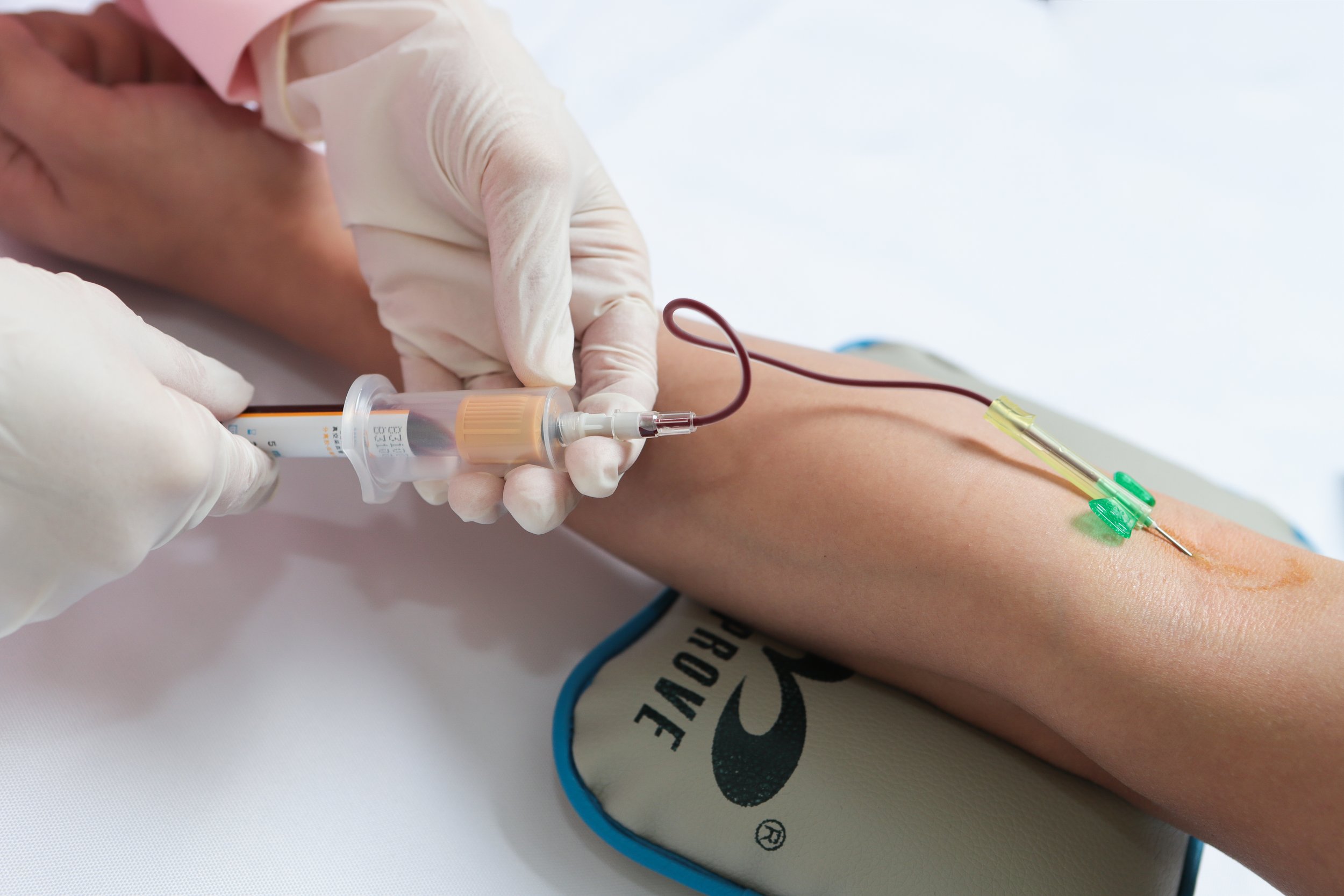
In a medical laboratory setting, enzyme-linked immunosorbent assay (ELISA) is a widely used technique for detecting the presence of antibodies or antigens in a patient's blood sample. This diagnostic test is crucial for diagnosing various Infectious Diseases, autoimmune disorders, allergies, and even some types of cancer. In this article, we will outline the key steps involved in performing an ELISA test in a medical laboratory setting, highlighting the importance of accuracy and precision in obtaining reliable diagnostic results.
Step 1: Coating the Plate
The first step in performing an ELISA test is coating the wells of a microtiter plate with the antigen of interest. This antigen can be a specific protein, peptide, or other molecules that will interact with the antibodies present in the patient's blood sample. The plate is typically coated overnight at 4°C to ensure proper adhesion of the antigen to the wells.
Key Points:
- Use a specific concentration of antigen to coat the plate.
- Incubate the plate at 4°C to facilitate antigen binding.
Step 2: Blocking Non-Specific Binding Sites
After coating the plate with the antigen, the next step is to block any non-specific binding sites to prevent false-positive results. This is done by adding a blocking agent, such as bovine serum albumin (BSA) or non-fat dry milk, to the wells and incubating the plate for a specific period of time. Blocking agents help reduce background noise and increase the specificity of the assay.
Key Points:
- Choose a suitable blocking agent based on the assay requirements.
- Optimize the blocking step to minimize non-specific binding.
Step 3: Adding the Sample
Once the plate is coated and blocked, the next step is to add the patient's blood sample to the wells. The sample is diluted in a specific buffer to ensure proper interaction with the antigen. After adding the sample, the plate is incubated to allow the antibodies present in the sample to bind to the coated antigen.
Key Points:
- Dilute the sample according to the assay specifications.
- Incubate the plate to allow antibody-antigen binding.
Step 4: Washing the Plate
After the sample has been incubated, the plate is washed multiple times to remove any unbound antibodies or other components. Washing steps are crucial for removing debris and reducing background noise in the assay. Proper washing ensures the accuracy and specificity of the results obtained from the ELISA test.
Key Points:
- Use an appropriate buffer for washing the plate.
- Perform multiple washes to remove unbound components.
Step 5: Adding the Detection Antibody
Following the washing step, a detection antibody is added to the wells to bind to the antibody-antigen complex formed in the previous steps. The detection antibody is typically conjugated with an enzyme, such as horseradish peroxidase (HRP) or alkaline phosphatase, which will produce a signal in the presence of a substrate. The enzyme-substrate reaction generates a colorimetric or chemiluminescent signal that can be measured and quantified.
Key Points:
- Select a suitable detection antibody for the target antigen.
- Choose an enzyme substrate that produces a detectable signal.
Step 6: Reading the Results
Once the detection antibody has been added and incubated, the final step is to read and interpret the results of the ELISA test. The colorimetric or chemiluminescent signal generated by the enzyme-substrate reaction is quantified using a spectrophotometer or luminometer. The intensity of the signal is directly proportional to the concentration of antibodies present in the patient's sample.
Key Points:
- Measure the absorbance or luminescence of each well using appropriate instrumentation.
- Compare the results to a standard curve to determine antibody concentration.
Conclusion
Performing an ELISA test in a medical laboratory setting involves several key steps that are essential for obtaining reliable and accurate diagnostic results. From coating the plate with the antigen to reading and interpreting the results, each step plays a crucial role in ensuring the specificity and sensitivity of the assay. By following the proper protocols and optimizing each step, Healthcare Providers can rely on ELISA tests to diagnose various diseases and monitor patient responses to treatment effectively.
Disclaimer: The content provided on this blog is for informational purposes only, reflecting the personal opinions and insights of the author(s) on the topics. The information provided should not be used for diagnosing or treating a health problem or disease, and those seeking personal medical advice should consult with a licensed physician. Always seek the advice of your doctor or other qualified health provider regarding a medical condition. Never disregard professional medical advice or delay in seeking it because of something you have read on this website. If you think you may have a medical emergency, call 911 or go to the nearest emergency room immediately. No physician-patient relationship is created by this web site or its use. No contributors to this web site make any representations, express or implied, with respect to the information provided herein or to its use. While we strive to share accurate and up-to-date information, we cannot guarantee the completeness, reliability, or accuracy of the content. The blog may also include links to external websites and resources for the convenience of our readers. Please note that linking to other sites does not imply endorsement of their content, practices, or services by us. Readers should use their discretion and judgment while exploring any external links and resources mentioned on this blog.