
Histology and Cytology Techniques in Medical Laboratories: A Comprehensive Overview
Summary
- Histology and cytology are essential techniques used in medical labs to analyze tissue and cellular specimens.
- Specific techniques like staining, microscopy, and cell culture are commonly used in histology and cytology.
- These techniques help pathologists and lab technicians diagnose various diseases and conditions accurately.
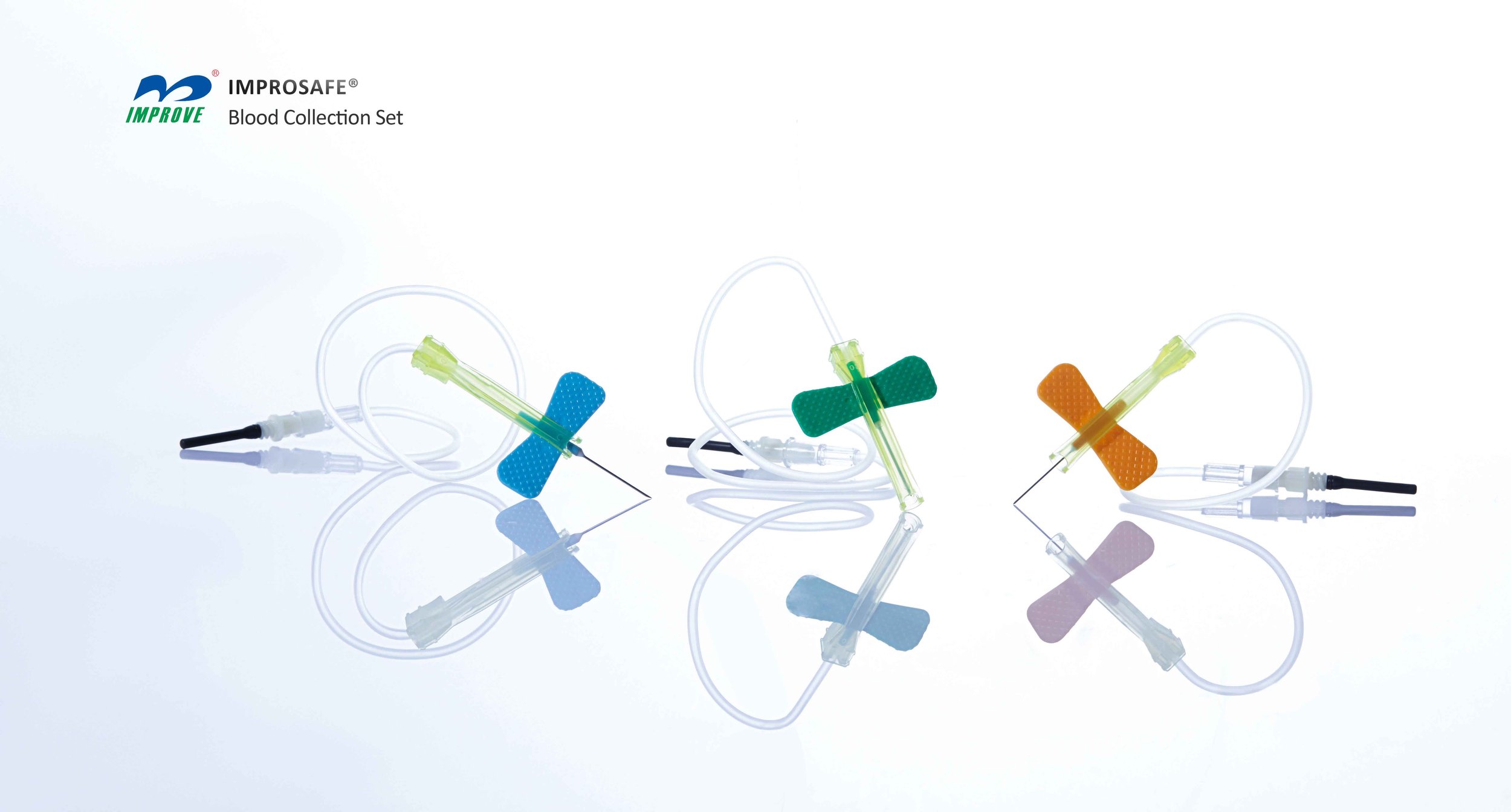
- Venipuncture: Phlebotomists use a needle to puncture a vein, usually in the arm, to collect blood samples for various tests.
- Skin puncture: In some cases, phlebotomists may collect blood samples by pricking the skin, typically on the finger or heel, for tests like glucose monitoring in diabetic patients.
- Specimen Collection: Phlebotomists follow strict protocols to collect and handle specimens safely to prevent contamination and ensure accurate Test Results.
Introduction
Medical laboratories play a crucial role in the diagnosis and treatment of diseases by analyzing various types of specimens. Histology and cytology are two important techniques used in these labs to examine tissue and cellular samples. In the United States, trained professionals, known as phlebotomists, use specific techniques to analyze and interpret these samples accurately.
Techniques Used in Histology
Staining
One of the primary techniques used in histology is staining, where tissue samples are treated with dyes to highlight specific structures or proteins. This helps pathologists visualize different cells and tissues under a microscope. Common stains used in histology include hematoxylin and eosin (H&E), which highlight cell nuclei and cytoplasm, and periodic acid-Schiff (PAS), which is used to detect carbohydrates in tissues.
Microscopy
Microscopy is another essential technique in histology, where pathologists examine stained tissue samples under a microscope. This allows them to identify abnormalities, such as cancer cells or infections, in the tissue. Electron microscopy is a specialized form of microscopy that provides high-resolution images of cellular structures, aiding in the diagnosis of rare diseases or genetic disorders.
Cell Culture
Cell culture is a technique used in histology to grow and maintain cells in a controlled environment. This is particularly useful in studying the behavior of cells in response to different stimuli or treatments. Cell culture techniques help researchers understand the molecular mechanisms underlying diseases like cancer and develop targeted therapies.
Techniques Used in Cytology
Cellular Smear
In cytology, a cellular smear is prepared by spreading a thin layer of cells on a glass slide, which is then stained and examined under a microscope. This technique is commonly used to analyze cells from body fluids like blood or sputum. Cellular smears help in the early detection of cancerous or pre-cancerous cells in the body.
Cell Block Preparation
Cell block preparation is a technique used in cytology to concentrate cells from fluid samples into a solid block for histological examination. This allows pathologists to evaluate the cellular architecture and characteristics of the sample more effectively. Cell block preparation is often used in the diagnosis of tumors or Infectious Diseases.
Flow Cytometry
Flow cytometry is a modern technique used in cytology to analyze the physical and chemical characteristics of cells. This technique involves passing cells through a laser beam and detecting the scattered light to identify different cell types. Flow cytometry is used in diagnosing blood cancers, immune system disorders, and monitoring the effectiveness of treatments.
Role of Phlebotomists in Medical Labs
Phlebotomists play a crucial role in collecting blood and other specimens for analysis in medical laboratories. They are trained to perform Venipuncture and skin punctures to obtain blood samples from patients. Phlebotomists also ensure that specimens are properly labeled, stored, and transported to the lab for analysis.
Phlebotomy Techniques
Conclusion
Histology and cytology are essential techniques used in medical laboratories to analyze tissue and cellular specimens accurately. By employing staining, microscopy, cell culture, cellular smears, cell block preparation, and flow cytometry, pathologists and lab technicians can diagnose various diseases and conditions with precision. Phlebotomists play a crucial role in collecting blood and other specimens for analysis, ensuring that accurate Test Results are obtained for patient care.
Disclaimer: The content provided on this blog is for informational purposes only, reflecting the personal opinions and insights of the author(s) on the topics. The information provided should not be used for diagnosing or treating a health problem or disease, and those seeking personal medical advice should consult with a licensed physician. Always seek the advice of your doctor or other qualified health provider regarding a medical condition. Never disregard professional medical advice or delay in seeking it because of something you have read on this website. If you think you may have a medical emergency, call 911 or go to the nearest emergency room immediately. No physician-patient relationship is created by this web site or its use. No contributors to this web site make any representations, express or implied, with respect to the information provided herein or to its use. While we strive to share accurate and up-to-date information, we cannot guarantee the completeness, reliability, or accuracy of the content. The blog may also include links to external websites and resources for the convenience of our readers. Please note that linking to other sites does not imply endorsement of their content, practices, or services by us. Readers should use their discretion and judgment while exploring any external links and resources mentioned on this blog.