
Quantitative Analysis Methods for Blood Specimens in Medical Labs
Summary
- Immunoassays and spectrophotometry are common methods used to perform quantitative analysis for blood specimens in medical labs.
- Flow cytometry and mass spectrometry are also utilized for more specialized testing.
- Accurate and precise quantification of blood components is essential for diagnosing and monitoring various health conditions.
Introduction
Medical laboratories play a crucial role in healthcare by providing accurate and reliable diagnostic information. Quantitative analysis of blood specimens is a common practice in medical labs to measure the levels of various components in the blood. This information is essential for diagnosing diseases, monitoring treatment efficacy, and assessing overall health status. In this article, we will explore the common methods used in medical labs to perform quantitative analysis for blood specimens in the United States.
Immunoassays
Immunoassays are widely used in medical laboratories to measure the concentration of specific proteins, hormones, and other substances in the blood. This method relies on the principle of antigen-antibody interactions to accurately quantify the target analyte. There are different types of immunoassays, including enzyme-linked immunosorbent assay (ELISA), radioimmunoassay (RIA), and chemiluminescent immunoassay (CLIA).
Steps involved in an immunoassay:
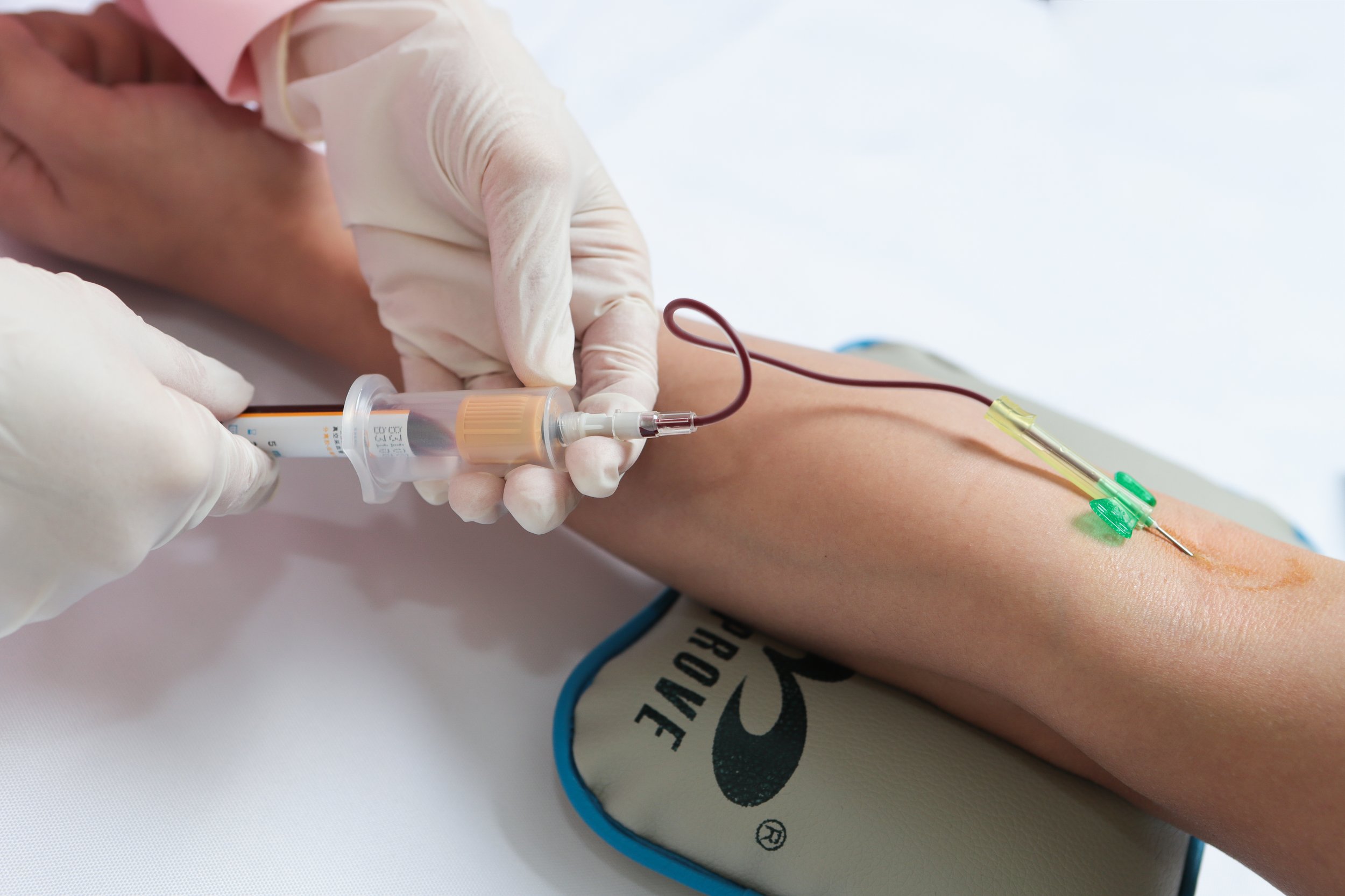
- Sample collection: Blood specimen is collected using Venipuncture or fingerstick.
- Sample preparation: The blood is centrifuged to separate the serum or plasma from the cellular components.
- Incubation: The sample is incubated with specific antibodies that bind to the target analyte.
- Washing: Unbound substances are washed away to reduce background interference.
- Detection: The amount of bound analyte is measured using colorimetric, luminescent, or radioactive signals.
- Quantification: The concentration of the analyte is determined based on the signal intensity.
Spectrophotometry
Spectrophotometry is a quantitative analysis technique that measures the amount of light absorbed or transmitted by a substance in the blood. This method is commonly used to determine the concentration of hemoglobin, glucose, cholesterol, and other blood components. Spectrophotometers are sophisticated instruments that can analyze the optical properties of a sample with high precision.
Principle of spectrophotometry:
- Beer-Lambert law: The absorbance of light by a substance is directly proportional to its concentration.
- Wavelength selection: Specific wavelengths of light are used to target the absorbance peak of the analyte.
- Calibration curve: Standard solutions with known concentrations are used to create a calibration curve for quantification.
- Data analysis: The absorbance readings are compared to the calibration curve to determine the concentration of the analyte in the sample.
Flow Cytometry
Flow cytometry is a specialized method used in medical labs to analyze and quantify individual cells in a heterogeneous population. This technique is commonly used for immunophenotyping, cell cycle analysis, and DNA content measurement. Flow cytometers can analyze thousands of cells per second and provide detailed information about cell size, morphology, and surface markers.
Workflow of flow cytometry:
- Sample preparation: Blood cells are stained with fluorescent antibodies or dyes that bind to specific cell markers.
- Fluidics system: The sample is injected into a sheath fluid to form a single-cell stream for analysis.
- Laser excitation: Cells pass through a laser beam that excites the fluorophores, emitting fluorescent light.
- Detection system: Detectors capture the emitted light signals and convert them into electronic signals.
- Data analysis: Software analyzes the fluorescence intensity and generates quantitative data on cell populations.
Mass Spectrometry
Mass spectrometry is a powerful analytical tool used in medical labs for quantitative analysis of small molecules, peptides, and proteins in blood specimens. This method can accurately measure the mass-to-charge ratio of ions in a sample, allowing for identification and quantification of analytes with high sensitivity and specificity. Mass spectrometers are equipped with sophisticated detectors and software for data analysis.
Steps involved in mass spectrometry:
- Ionization: The sample is ionized to generate charged particles for analysis.
- Mass analysis: The ions are separated based on their mass-to-charge ratio in a mass analyzer.
- Detector: The ions are detected by a sensitive detector that converts the signals into electronic data.
- Data processing: Software analyzes the mass spectra and calculates the abundance of different ions in the sample.
- Quantification: The concentration of the analyte is determined by comparing the peak intensities to internal standards or calibration curves.
Conclusion
Quantitative analysis of blood specimens is essential for diagnosing and monitoring various health conditions in patients. Immunoassays, spectrophotometry, flow cytometry, and mass spectrometry are common methods used in medical labs to accurately measure the levels of different components in the blood. These techniques provide valuable information for Healthcare Providers to make informed decisions about patient care. Continued advancements in laboratory technology and methodology will further enhance the accuracy and reliability of quantitative analysis in medical labs.
Disclaimer: The content provided on this blog is for informational purposes only, reflecting the personal opinions and insights of the author(s) on the topics. The information provided should not be used for diagnosing or treating a health problem or disease, and those seeking personal medical advice should consult with a licensed physician. Always seek the advice of your doctor or other qualified health provider regarding a medical condition. Never disregard professional medical advice or delay in seeking it because of something you have read on this website. If you think you may have a medical emergency, call 911 or go to the nearest emergency room immediately. No physician-patient relationship is created by this web site or its use. No contributors to this web site make any representations, express or implied, with respect to the information provided herein or to its use. While we strive to share accurate and up-to-date information, we cannot guarantee the completeness, reliability, or accuracy of the content. The blog may also include links to external websites and resources for the convenience of our readers. Please note that linking to other sites does not imply endorsement of their content, practices, or services by us. Readers should use their discretion and judgment while exploring any external links and resources mentioned on this blog.